Description
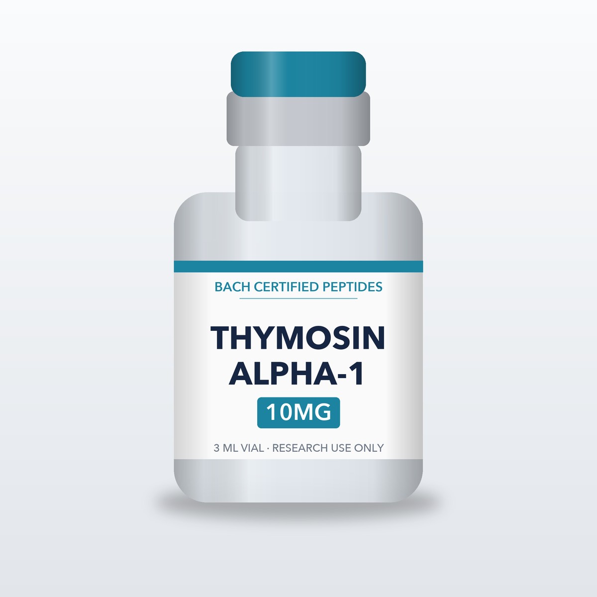
| THYMOSIN ALPHA-1 10MG | |
|---|---|
| SKU | CP_THYMOSIN_ALPHA_1_10MG_3ML_VIAL |
| CATEGORY | Research Compound |
| CAS | 62304-98-7 |
| VIAL SIZE | 3 mL |
| FORM | LYOPHILIZED POWDER |
| MOLECULAR WEIGHT | 3108.32 g/mol |
| SEQUENCE | Ser-Asp-Ala-Ala-Val-Asp-Thr-Ser-Ser-Glu-Ile-Thr-Thr-Lys-Asp-Leu-Lys-Glu-Lys-Lys-Glu-Val-Val-Glu-Glu-Ala-Glu-Asn |
| MOLECULAR FORMULA | C129H215N33O55 |
| ITEM ID | 845 |
THYMOSIN ALPHA-1 10MG provides Thymosin alpha-1 as lyophilized powder in a 3 mL vial.
Research Context
Thymosin alpha-1 has been investigated in nonclinical models across immunology, oncology, and neuroscience. Below are summaries of nonclinical findings drawn from the supplied literature.
In Vitro CD8⁺ T-Cell Studies
Mishra et al. (2026) examined Tα1 effects on human CD8⁺ T-cells cultured under four conditions: untreated, CD3/CD28-stimulated, Tα1-treated, and combined CD3/CD28 plus Tα1. Using CFSE-based flow cytometry, the investigators measured proliferation indices and surface expression of activation markers (CD69, CD25, HLA-DR) and exhaustion markers (PD-1, TIM-3, LAG-3). The combination condition showed increased proliferation index and elevated activation-marker expression relative to individual treatments. In a chronic-stimulation model, Tα1 treatment was associated with reduced expression of PD-1, TIM-3, and LAG-3 on CD8⁺ T-cells. Cytokine secretion (IL-2, IFN-γ, TNF-α, IL-10) was measured by multiplex bead-based assay and was elevated in the combination group [1].
Dendritic Cell Signaling and Chemotherapy-Derived Apoptotic Bodies (Murine Model)
Wei et al. (2026) investigated Tα1 interactions with apoptotic bodies (ABs) released from chemotherapy-treated tumor cells in murine models. The study reported that Tα1 bound to tumor ABs and interacted with AB-borne miR-146a-5p within the endolysosomal compartment of dendritic cells (DCs). This interaction was associated with reduced lysosomal RNase A-mediated degradation of miR-146a-5p, facilitating TLR7 activation. Downstream readouts included DC maturation markers, migration to tumor-draining lymph nodes, and CD8⁺ T-cell activation assays. The authors described TLR7-dependent effects in tumor-bearing mice with high miR-146a-5p expression [2].
HCRTR1 Engagement in Murine Ischemic Stroke Model
Kang et al. (2026) reported that Tα1 acts as a ligand for Hypocretin (Orexin) Receptor 1 (HCRTR1) on neurons. In murine ischemic stroke models, genetic deletion of the Tα1-encoding gene Ptma was associated with increased injury markers, while Tα1 administration was associated with reduced RIPK3-mediated necroptosis signaling. The study measured circulating Tα1 levels, neuronal cell-death markers, and behavioral endpoints in mice [3].


Reviews
There are no reviews yet.